Order
Contacts

Clarus Fundus Photography
Clarus Fundus Photography — What It Is and Why We Do It
Clarus Fundus Photography is a high-resolution photo of the back of your eye (the retina), taken with a special camera. This isn’t just a regular picture — it shows the tiny structures that make vision possible, including the retina, optic nerve, blood vessels, and macula. These detailed images help your doctor understand your eye health better than what can be seen with just a routine exam or simple eye check.
What This Test Does
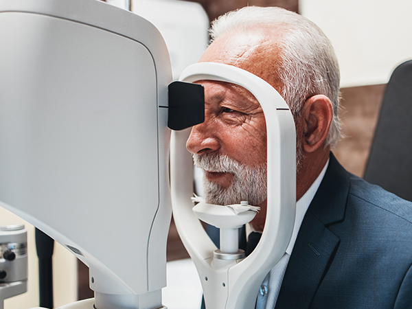
During the test, you’ll place your chin and forehead on a support and look into a camera. The Clarus system captures a true-color, ultra-wideview image of the retina — often showing a larger area than traditional cameras — so your doctor can see both the central and peripheral parts of your retina clearly.These photos look very similar to what your eye doctor sees when they examine your retina in person, which makes them especially helpful for diagnosing and tracking eye disease over time.
Why We Do This Test
We use Clarus Fundus Photography because it helps your doctor:Detect eye diseases early — many serious eye conditions develop without symptoms at first.
Document the appearance of your retina so changes can be compared from year to year.
Monitor disease progression or response to treatment.
Guide treatment decisions with detailed visual information.
Who Might Need This Test
Your eye doctor may recommend Clarus Fundus Photography if you have conditions that can affect the retina or optic nerve, including:
Diabetes or high blood pressure — to check for retinal blood vessel damage.
Age-related macular degeneration (AMD) — to look for early changes in the macula.
Glaucoma — to monitor the optic nerve and retinal nerve fiber layer.
Retinal tears, detachments, or peripheral changes — which can occur without warning.
Visual symptoms such as flashes, floaters, or unexplained vision changes — to investigate the cause.
In Summary
Clarus Fundus Photography gives your doctor a detailed, wide-angle picture of the back of your eye, helping detect, evaluate, and manage eye conditions sooner and more effectively. It’s a key part of comprehensive eye care that supports long-term vision health.

Our Locations
Yankton, SD
415 W. 3rd St. Yankton, SD 57078
415 W. 3rd St. Yankton, SD 57078
Hartington, NE202 S. Robinson Ave. Hartington, NE 68739
Creighton, NE
817 N. Main St.
Creighton, NE 68729
817 N. Main St.
Creighton, NE 68729
OFFICE HOURS
- Monday8:00AM — 5:00PM
- Tuesday8:00AM — 5:00PM
- Wednesday8:00AM — 5:00PM
- Thursday8:00AM — 5:00PM
- Friday8:00AM — 2:00PM
- SaturdayClosed
- SundayClosed
© 2026 Willcockson Eye Associates. All rights Reserved. Accessibility Statement - Privacy Policy - Sitemap
Managed and Designed By